How Does the Heart Work? Chambers, Valves and the Electricity Behind Every Beat
Your heart beats about 100,000 times a day. How its four chambers, four valves and built-in electrical system move blood around your body.
Short answer
The heart is a muscular pump about the size of a fist that moves blood around the body in two loops. Its right side receives oxygen-poor blood from the body and pumps it to the lungs, where it picks up oxygen. Its left side receives this oxygen-rich blood from the lungs and pumps it out through the aorta to the rest of the body. Each side has an upper chamber, the atrium, and a lower, more muscular chamber, the ventricle, and four valves keep blood flowing in one direction. Every beat starts with an electrical signal from the heart’s natural pacemaker, the sinoatrial node, so the heart keeps beating on its own, about 60 to 100 times a minute at rest.
In this article (8)
Put your hand on your chest and you can feel it: a steady rhythm that has not stopped once since before you were born. Your heart beats roughly 100,000 times a day and more than 2.5 billion times in an average lifetime, without a single conscious command. It is a pump, an electrical system and a set of precision valves, all packed into an organ about the size of your fist.
A double pump
The heart sits in the middle of the chest, behind the breastbone and between the lungs, tilted slightly to the left. It is really two pumps side by side, separated by a muscular wall called the septum:
- The right side pumps blood to the lungs.
- The left side pumps blood to the rest of the body.
Each side has two chambers. The upper chamber, the atrium, collects blood coming in. The lower chamber, the ventricle, is thicker and more muscular and pushes blood out. The left ventricle has the thickest wall of all, because it must push blood all the way to your toes and brain.
The journey of a drop of blood
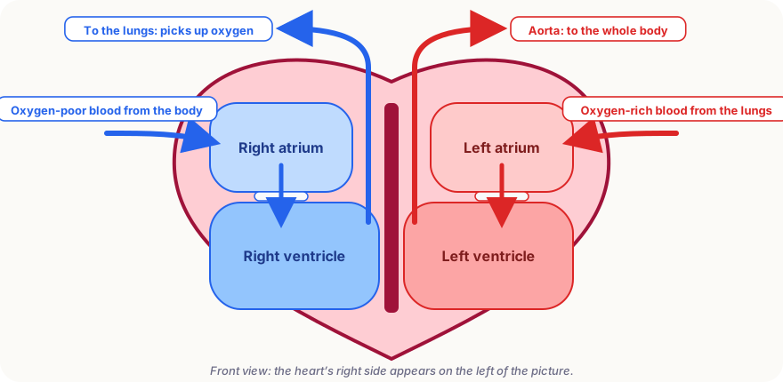
According to the US National Heart, Lung, and Blood Institute (NHLBI), blood travels through the heart in a fixed order:
- Oxygen-poor blood returns from the body through two large veins into the right atrium.
- It passes through the tricuspid valve into the right ventricle.
- The right ventricle pumps it through the pulmonary valve to the lungs, where it releases carbon dioxide and picks up oxygen.
- Oxygen-rich blood returns to the left atrium.
- It passes through the mitral valve into the left ventricle.
- The left ventricle pumps it through the aortic valve into the aorta, the body’s largest artery, and from there to every organ.
This creates two circuits: a short pulmonary circulation through the lungs, and a long systemic circulation through the body. At rest, the heart moves around 5 litres of blood a minute, roughly the whole volume of blood in an adult body.
| Part | Job |
|---|---|
| Right atrium | Receives oxygen-poor blood from the body |
| Right ventricle | Pumps blood to the lungs |
| Left atrium | Receives oxygen-rich blood from the lungs |
| Left ventricle | Pumps blood to the whole body (thickest wall) |
| Tricuspid and mitral valves | Stop blood flowing back into the atria |
| Pulmonary and aortic valves | Stop blood flowing back into the ventricles |
| Coronary arteries | Supply the heart muscle itself with blood |
Valves: one-way doors
The four valves are thin, strong flaps of tissue that open in one direction only. They open to let blood through and snap shut to stop it flowing backwards. That snapping is what a doctor hears through a stethoscope. According to the NHLBI, the familiar “lub-DUB” comes from two sets of valves closing: “lub” when the valves between atria and ventricles close, and “dub” when the aortic and pulmonary valves close.
When a valve does not close properly, some blood leaks backwards, creating an extra sound called a murmur. Many murmurs are harmless, but some point to valve problems that need treatment.
The electricity behind every beat
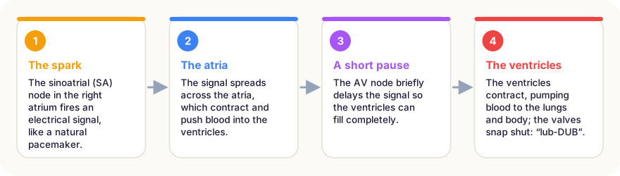
Unlike the muscles in your arms, the heart does not wait for instructions from the brain. It has its own electrical system:
- A small cluster of cells in the right atrium, the sinoatrial (SA) node, fires an electrical signal. It is the heart’s natural pacemaker.
- The signal spreads across both atria, which contract and push blood down into the ventricles.
- At the atrioventricular (AV) node, the signal pauses for a fraction of a second, giving the ventricles time to fill.
- The signal then races down special fibres into the ventricles, which contract powerfully from the bottom up, squeezing blood out like toothpaste from a tube.
The brain does influence the heart: nerves and hormones such as adrenaline speed it up during exercise or fear and slow it down during rest. But the basic rhythm comes from the heart itself. An electrocardiogram (ECG) records these electrical signals from the skin, and doctors use it to spot rhythm problems.
The heart in numbers
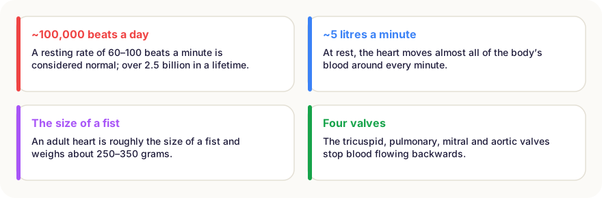
A normal resting heart rate for adults is generally 60 to 100 beats per minute. During hard exercise it can more than double, and the amount of blood pumped each minute can rise several times over. Trained athletes often have resting rates below 60, because their stronger hearts pump more blood with each beat.
Blood pressure measures the force of blood against artery walls. The first number (systolic) is the pressure when the ventricles contract; the second (diastolic) is the pressure between beats.
Feeding the pump
Although the heart is full of blood, its muscle cannot absorb oxygen from the blood inside its chambers. It has its own supply: the coronary arteries, which branch off the aorta and wrap around the heart. When one of these arteries becomes blocked, part of the heart muscle is starved of oxygen; this is a heart attack. Chest pain or pressure, shortness of breath and pain spreading to the arm or jaw are warning signs that need emergency help immediately.
Keeping your heart healthy
Heart disease is one of the leading causes of death worldwide, but much of the risk can be reduced. Regular physical activity, not smoking, a balanced diet with plenty of vegetables and whole grains, a healthy weight, enough sleep and keeping blood pressure, cholesterol and blood sugar under control all help the heart do its lifelong job.
In short
- The heart is a double pump: the right side sends blood to the lungs, the left side to the body.
- It has four chambers (two atria, two ventricles) and four valves that keep blood flowing one way.
- The “lub-dub” sound is the valves closing.
- Each beat starts in the sinoatrial node, the heart’s built-in pacemaker.
- At rest the heart beats about 60–100 times a minute and pumps about 5 litres of blood a minute.
- The heart muscle gets its own blood through the coronary arteries.
Frequently asked questions
Which side of the body is the heart on?
The heart sits in the middle of the chest, behind the breastbone, tilted slightly to the left. Its strong left ventricle points down and to the left, which is why the heartbeat is easiest to feel on the left.
What is a normal resting heart rate?
For adults, about 60 to 100 beats per minute is generally considered normal at rest. Fit athletes often have lower rates. If your heart rate seems unusual or you have symptoms, talk to a doctor.
What makes the ‘lub-dub’ sound?
The heart sounds come from valves closing. ‘Lub’ is the valves between the atria and ventricles snapping shut; ‘dub’ is the aortic and pulmonary valves closing after the ventricles have pumped.
Does the heart ever rest?
It rests only in tiny pauses between beats. Heart muscle is built to work continuously and has a very rich blood supply of its own, through the coronary arteries, to keep it going for a lifetime.
Sources
- National Heart, Lung, and Blood Institute (NIH) – How the Heart Works
- National Heart, Lung, and Blood Institute (NIH) – How the Heart Beats
- National Heart, Lung, and Blood Institute (NIH) – Blood Flow
This article is based on peer-reviewed research and publications from trusted institutions, and it is reviewed regularly. How we work →
You might also wonder
All in Human Body →
What Are Blood Types? A, B, AB, O and Rh Explained, Plus Who Can Donate to Whom
Blood types depend on tiny markers on red blood cells. What A, B, AB, O and Rh mean, how blood types are inherited and who can give blood to whom.
How Does the Immune System Work? Your Body’s Defence Force, Explained Simply
Your immune system uses barriers, fast responders and cells that learn and remember. How it fights germs, how vaccines train it and what goes wrong.

Why Do Knuckles Crack? The Bubble Behind the Pop, and Whether It Causes Arthritis
Cracking your knuckles forms a gas cavity in the joint. What MRI scans revealed, why the same knuckle won’t crack twice in a row and whether it is harmful.
Why Do We Need Sleep? What Happens in Your Brain and Body Every Night
We spend about a third of our lives asleep. What sleep does for memory, the brain and immunity, how much you need at every age and what happens without it.

Why Do We Blush? The Science of a Very Human Reaction
Blushing is the only facial expression we cannot fake or hide. What happens in the body when we turn red, why does it start, and is it actually useful?
How Does Caffeine Keep You Awake? Adenosine, Half-Life and Sleep
Caffeine doesn’t give you energy; it blocks a sleepiness signal in the brain. How it works, how long it lasts, how much is safe and why it can ruin your sleep.